Feline Streptococcal (G-Strep)
Cat G-Strep
"G-Strep" is the short version of "Group G Streptococcal". "G-Strep" is part of the normal microbial flora of the gastrointestinal tract, vagina, and skin of cats. "Streptococci" are normally non-pathogenic, but a few species can cause significant morbidity and mortality ranging from death in young kittens, from pyometra, pneumonia and upper respiratory infection.
Gallery
Click on each picture for details and credit information.


Symptoms
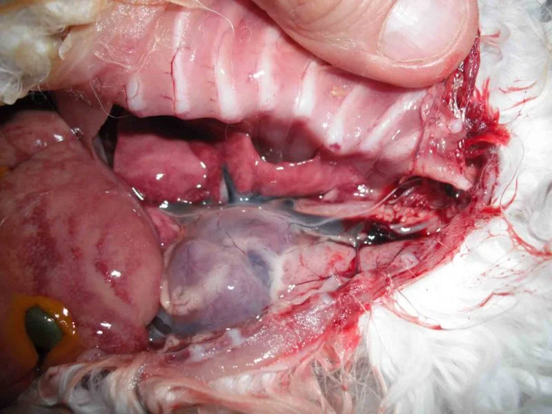
"Streptococci Infections" in adult cats have a high probability of show no symptoms! Unfortunately kittens have a weak immune system making them more subseptable to death may show symptoms prior to death. Many kittens will show no clinical symptoms of illness until hours before death. Usually not all of the kittens in the litter are affected. Kittens are typically infected with the bacteria from the queen's vagina during birth. This can result in an abscess of the umbilicus which spreads into the liver and abdominal cavity, leading to peritonitis and septicaemia. Some kittens may have an obvious umbilical abscess or swelling.
Unexplained spontaneous abortions or absorption.
Unexplained death of newborn kittens.
Birth abnormalities (Intestines on outside, cleft palette).
Affected kittens die between 1 and 10 days, rare cases of up to 6 weeks old.
No clinical symptoms of illness until hours before death.
Affected kittens show slower than average weight gain.
All the signs of Chlamydia but negative on testing.
Difficulty swallowing due to swelling.
Fever
Lethargy
Coughing
Pneumonia
Concerns
Dehydration: Re-hydration is also important for restoring the body with fluid and flushing the system of the infection. Kittens will be unable to digest food if too dehydrated.
Weight-loss: All felines, especially kittens, in your household should be weighed immediately so you can determine weightloss.
Respiratory Distress: Cat(s) showing signs of respiratory distress of any level should be seen by the veterinarian immediately they may require intensive veterinary supportive care (Incubator with oxygen therapy, fluids and antibiotics).
Diagnosis
G-strep requires specialized equipment to grow however other common bacteria can be detected through vaginal smear with sensitivity test. Many veterinarians are not aware of G-Strep and can mistake it as Upper Respiratory Infection, Chlamydia, Calicivirus or Panleukopenia.
Highly suggested to perform a feline upper respiratory PCR test and bacterial culture to rule out other possible ailments that present with the same symptoms such as Feline Mycoplasma.
Vaginal culture with sensitivity test on a female if you suspect G-Strep (must been done with the female is in heat). G-strep may not be detected even if infected.
Your culture will take about 10 days, and will most likely come back with "normal flora", because the G Strep is at such a low level that it won't "register" on the lab tests.
Cases have been documented can ecolli imbalance found through vaginal smear. This makes testing crucial to determine if breeders are treating for the wrong problem!
Treatment
Consult with your veterinarian before any treatment.
DO NOT USE GENERIC BRAND MEDICATIONS
Beta-haemolytic Streptococci are usually resistant to sulfamethoxazole, tetracycline and gentamicin. Resistance to penicillins or cephalosporins is very rarely detected.
Penicillin G
Penicillin and other beta-lactam antibiotics are generally effective against GGS infections. Penicillin is often the first choice due to its narrow spectrum of activity, cost-effectiveness, and few adverse effects. For those with penicillin allergies, other options like clindamycin, clarithromycin, or azithromycin are recommended.
Group G Streptococcal (GGS) infections: They can cause a wide range of infections, from mild skin and soft tissue infections to more serious invasive diseases like bacteremia, endocarditis, and septic arthritis. GGS is often found as part of the normal flora in the pharynx, gastrointestinal tract, genital tract, and skin, but it can also cause opportunistic infections in individuals with underlying medical conditions.
Clindamycin
After clindamycin treatment, the G Strep will most likely still be retained in the normal flora at a very low level. Therefore, the cat could always be a potential G Strep carrier with respect to breeding only. In other words, we should be concerned with the breeding aspects of the Strep G rather than whether the cats do not have any G Strep at all, since the clindamycin will most likely not get every last bit of it - even at a 3 week dose.
UPDATES…..
“Please see the G Strep Updates: The Continuing Story: Part 2 and Additional Comments and Follow Up to G Strep article (2004). Read all three articles before treating your cats. Since I wrote my articles, I have received feedback and requests for information for literally several hundred breeders around the world. It seems that this problem is becoming even more widespread, in every country and in every breed.
https://www.7thheavencats.com/g-strep-1.html
https://www.7thheavencats.com/g-strep-2.html
https://www.7thheavencats.com/g-strep-3.html”
References
http://www.encyclopedia.com/topic/Streptococcal_infections.aspx
http://www.petmd.com/cat/conditions/respiratory/c_ct_streptococcal_infections
http://veterinarynews.dvm360.com/implications-emerging-streptococcal-diseases
http://www.life.umd.edu/classroom/bsci424/pathogendescriptions/Streptococcus
http://www.rasekatter.no/artikler/group_g_streptococcus_bacteria.php
Stories From Breeders
Please read the breeder original website posts!
“All was well until the kittens were ten days old. That morning, I picked up the first baby, to check them as I usually did, first thing when I woke up. The baby’s little nostrils were clogged shut with mucous! I had never seen signs of URIs (upper respiratory infection) in such young babies, so, alarmed, I quickly picked up the next kitten. All the babies had clogged nostrils and it seemed to me that their breathing was labored. Despite Baytril, Clavomox and Amoxi all kittens died one by one.
A girl (one of my DMs) who had had four healthy litters of 5-6 had a litter with five kittens, four of them stillborn. Only one lived.
A young girl I had been showing, with over 160 points to her grand, on the morning of a show, she presented herself to me with a pyometra. I rushed her to the veterinarian. She was treated successfully with Baytril and prostaglandins. I bred her on the next heat. The breeding took. She aborted her kittens at eight weeks. I bred her again. It took. She AGAIN aborted her kittens at eight weeks into the pregnancy.
My other DM girl who had had two good litters was bred to my stud THREE times and the breeding did not take.
I had litters of one or two kittens where before I had had normal litters of 4-6. In two of these litters of one, the kitten was fine in the morning and suddenly dead in the evening - signs of a quick and intense pneumonia and nothing I could do.
A girl who came for breeding (all tests and healthy) produced two kittens with spinal deformities that died at a few weeks of age. The sire had had many healthy litters with never any sort of deformity or problem.
Needless to say, I couldn’t understand what was going on as all blood panels and other diagnostic tests came back totally normal, there were no signs of ill health: coats were shiny, eyes were bright, appetites were excellent, energy levels were great. I was getting really discouraged and depressed.
Then, a breakthrough.. A breeder friend in Australia forwarded me an e-mail from an Australian feline health list. The post was from Dr. Sue Rodger-Withers PhD, a microbiologist and university lecturer in that country. She and her colleagues had done research on low grade uterine infections due to Gp G-Strep (Group G Streptococcus bacteria) and the use of Antirobe (clindamycin) for treatment.
Read more: https://www.rasekatter.no/artikler/group_g_streptococcus_bacteria.php”
Feline Mycoplasma
Feline Mycoplasma
Mycoplasma species are small bacteria that lack a peptidoglycan cell wall. They belong to the normal commensal flora of the conjunctiva and upper airways (pharynx, larynx, oral cavity, nasal cavity) in cats and are a well-recognized cause of conjunctivitis and upper respiratory infection in this species. This disease can cause death if not diagnosed and treated by a veterinarian. Over 100 species have been included in the genus Mycoplasma. Microbes of the class Mollicutes, to which Mycoplasma belongs, are parasites or commensals of humans, animals, and plants. The genus Mycoplasma uses vertebrate and arthropod hosts. Dietary nitrogen availability has been shown to alter codon bias and genome evolution in Mycoplasma and Phytoplasma.
Symptoms
Anemia.
Lethargy.
Lack of appetite.
Weakness.
Fever.
Yellowing of skin (Jaundice)
Pale skin and mucous membranes.
Nasal or ocular (eye) discharge.
Weak newborns, stillbirths, early death of newborns, or death while in embryo.
Diagnosis
PCR Upper Respiratory Panel
https://www.idexx.com/en/veterinary/reference-laboratories/idexx-realpcr-tests/
https://tvmdl.tamu.edu/tests/feline-upper-respiratory-panel-qpcr/
Bacterial Culture (Sensitivity test suggested)
Treatment
Only Gram-Negative Antibiotics Are Effective
Doxycycline (Preferred treatment)
(34mg) per 5lbs of cat
Enrofloxacin
(5 mg/kg/day, PO) is a suitable alternative to doxycycline.
Enrofloxacin given at 5 mg/kg, PO, every 24 hours or at 10 mg/kg, PO, every 24 hours for 14 days is tolerated by cats and is
Equally effective or more effective than doxycycline
Azithromiacin
Albuterol sulfate (Repository Distress)
Prevention
Mycoplasma genitalium (mycoplasma) has recently been identified as a sexual transmitted infection and is a bacteria that infects the mucous membranes of the urethra,cervix, throat and anus. Mycoplasma is transmitted by vaginal, anal and oral sex.
“Bacterial infections and /or Mycoplasma in the semen (click for further info) - Overgrowth of bacteria and/or mycoplasma can adversely affect sperm morphology, motility and production. Semen cultures, when evaluated in conjunction with poor quality semen can diagnose pathogenic bacteria, mycoplasma and/or ureaplasma (we prefer Michigan State University’s test - Semen culture and sensitivity with added ureaplasma and mycoplasma -speciated). Beta strep, E-coli, Streptococcus, Pseudomonas and other bacteria in abundance should be treated by your veterinarian with the appropriate antibiotics . Mycoplasma Canis and Mycoplasma Cynos are pathogens that have been shown to significantly reduce fertility. All infections should be treated with appropriate antibiotics as soon as detected. Doxycycline is the best available choice currently available for Mycoplasma (currently needs to be compounded). Frequently a combination of antibiotics are needed to resolve many infections. Please contact your veterinarian for the appropriate treatment. Some Mycoplasma’s (not Canis or Cynos) and bacteria’s can be considered “Normal Flora” in the absence of fertility problems. ”
Resources
https://www.merckvetmanual.com/circulatory-system/blood-parasites/hemotropic-mycoplasmas#v3257810
http://citeseerx.ist.psu.edu/viewdoc/download?doi=10.1.1.1009.7562&rep=rep1&type=pdf
https://veterinary-practice.com/article/treatment-of-mycoplasma-spp-infections-in-cats
Panosteitis in Dogs
Panosteitis in Dogs
By Cheryl Yuill, DVM, MSc, CVH
Medical Conditions
What is panosteitis?
"Panosteitis is sometimes called growing pains."
Panosteitis is a painful inflammation of the outer surface or shaft of one or more long bones of the legs. It is sometimes called "growing pains." Panosteitis may occur in more than one bone at a time or may move around from area to area, cause a "shifting" lameness that goes from one bone or leg to another. The lameness tends to occur very suddenly and usually occurs spontaneously, or without a history of trauma or excessive exercise.
Are all dogs affected with this condition?
Panosteitis is a condition that affects young, rapidly growing dogs. Although it can occur in any breed of dog, larger breeds, such as German Shepherds (most common), Great Danes, Golden Retrievers, Labrador Retrievers, Rottweilers, Doberman Pinschers, and Basset Hounds, are more prone to this problem. Affected dogs are usually between 5 and 14 months of age, but the first symptoms may occur as early as 2 months of age or as late as 18 months of age. Males seem to be affected more often than females, although either sex can develop panosteitis. Affected dogs often have recurrent episodes of panosteitis until they reach 2 years of age, at which time it will spontaneously resolve.
What is the cause?
Panosteitis is a painful condition, and the pain is likely caused by increased pressure within the bone, and/or by stimulation of pain receptors in the periosteum, or outer, soft tissue lining of the bone. The underlying cause of panosteitis is unknown, but genetics, stress, infection, metabolism, or an autoimmune component may be factors. Since German Shepherds seem to be particularly predisposed to panosteitis, there may be a genetic component to the disease, at least in this breed. It has also been suggested that rapid growth and high-protein, high-calcium diets may predispose some dogs to this condition.
What are the clinical signs?
The typical symptom is a sudden, unexplained, painful lameness of one or more legs. The lameness may be mild or severe. The most common bone that is affected is the humerus (upper arm), but panosteitis may also be found in the radius and ulna (the foreleg), the femur (thigh) and/or the tibia (lower rear leg). The affected bone will be painful to the touch. Other symptoms such as fever, anorexia, lethargy, or weight loss may be noticed.
Panosteitis tends to have a cyclic nature, with periods of worsening symptoms followed by periods of improvement. The pain often shifts from leg to leg. Each episode of lameness may last for a few days to a few weeks, and the period between episodes is often about a month, but may vary.
How is it diagnosed?
When your veterinarian examines your dog, panosteitis will be suspected if the patient shows pain when pressure is applied to the affected bone(s). The diagnosis is confirmed by radiographs or x-rays, which usually show a characteristic increase in the density of the affected bones. The degree of change may not correlate to the severity of the lameness. In some cases, radiographic evidence may not be present for up to ten days after lameness begins; in these cases, repeat x-rays taken 2 weeks later will confirm the diagnosis. After the condition has resolved, the bone density normalizes and the bone looks normal on radiographs.
What is the treatment?
Although this disease is self-limiting, and will spontaneously resolve, during episodes of lameness the condition is very painful. At these times, treatment is supportive, using analgesics (pain medications) and/or anti-inflammatory drugs as needed.
"Pain control should always be given to help your pet feel more comfortable; denying your dog pain control is inhumane."
Pain control should always be given to help your pet feel more comfortable; denying your dog pain control is inhumane.
During episodes of lameness, exercise should be restricted. Between episodes, light to moderate exercise should be encouraged, but hard or vigorous exercise is discouraged, as are very long walks.
Some dogs with panosteitis have a poor appetite; in these cases, it is important to ensure that they are given a properly balanced and palatable diet. In some cases, supplements such as nutraceuticals, Omega fatty acids or antioxidants such as Vitamin C may be helpful.
What is the prognosis?
Panosteitis is a self-limiting disease, meaning that it will eventually go away. The disease should be completely resolved by the time the dog reaches 18-24 months of age. Each episode of lameness should last no longer than 3 weeks; if your pet's lameness persists without relief for longer than 4-5 weeks, it is likely that the dog is affected with another bone disorder (see our handout on Bone Diseases of Growing Dogs for further information).
If panosteitis is a self-limiting disease, why is it necessary to perform diagnostic procedures such as radiographs?
Although panosteitis is not a serious disease, and is a common cause of lameness, other, more serious bone diseases can cause lameness in young dogs. In order to be sure that a sudden onset of lameness is not caused by one of these more serious bone diseases, radiographs must be taken. If the radiographs show the typical lesions of panosteitis, then you can rest assured that your dog will eventually outgrow the problem.
Are there any preventive measures I can take to prevent panosteitis in my large breed dog?
"...it is contraindicated to feed large breed puppies with an adult dog food that contains lower levels of protein and calcium."
Although there are potential links between diets containing excessive levels of dietary protein and/or calcium, it is contraindicated to feed large breed puppies with an adult dog food that contains lower levels of protein and calcium. The reason for this is that adult dog food also has lower calories or energy levels than puppy food. Rapidly growing puppies require higher levels of dietary energy to meet the needs of growth, and will need to eat more of a low-energy food to meet these requirements. Eating more of a low energy diet will result in a higher overall intake of protein and calcium. A better option is to feed an affected dog a high quality diet that has been specifically formulated for use in large breed puppies or adolescents, and to restrict the quantity fed to keep the dog at a lean, healthy body weight. Do not allow your puppy to become overweight. Consult your veterinarian for further advice on the most appropriate nutrition for your dog.
This client information sheet is based on material written by: Cheryl Yuill, DVM, MSc, CVH
Kitten Incubator
Kitten Incubator
Sadly, Joe Freed, the owner of Petiatric, passed away. The entire rehabber community along with many breeders, are incredibly indebted to Joe for his kind heart and amazing work that will live on for years to come. I am now exclusively using Brinsea incubators (actually called “intensive care units”).
Brinsea incubators are great quality, easy to clean, and have solid sides to give the babies more security and reduce the stress on them. These units also have a digital temperature control, air filtration, an immersible bottom for easy cleaning, and a clear door so rehabbers can keep an eye on the babies.
You can find cheaper incubators on the market, and certainly homemade versions can be assembled for less, but they are not as reliable. Please consider Brinsea.
I own two of these!

Other Items You Might Need


Feline Upper Respiratory PRC Test
What is a PCR Test?
Polymerase chain reaction (PCR) is a process that enables the production of virtually unlimited copies of genetic material in the laboratory. PCR testing identifies a pathogen based on the presence of the pathogen’s DNA or RNA in the patient specimen, before antibody can be detected, making it a useful diagnostic tool for early disease detection in sick animals.
Bordetella bronchiseptica
Chlamydophila felis
Feline calicivirus (FCV)
Feline herpesvirus 1 (FHV-1; also known as feline rhinotracheitis virus)
Influenza
Mycoplasma felis
IDEXX RealPCR Tests
The IDEXX Canine Respiratory Disease (CRD) RealPCR Panel provides rapid, sensitive and specific identification for eight infectious agents—all at once. The IDEXX Feline Upper Respiratory Disease (URD) RealPCR Panel dramatically increases your chance of accurately identifying the origins of feline URD.
Canine code: 2524
Feline code: 2512
Equine code: 2740
https://www.idexx.com/en/veterinary/reference-laboratories/idexx-realpcr-tests/
https://www.zoologix.com/dogcat/Datasheets/FelineRespiratoryPanel.htm
Tube & Bottle Feeding Savannah Kittens
Tube & Bottle Feeding Kittens
Feeding Mixture
- Powder Mix (0-2 Weeks)
- Milk Matric Ratio is 1 scoop powder per 3 scoop water.
- Dry Mix (3-12 Weeks)
- Typical Ratio is 1 scoop dry, 1 extra scoop water per every 2 scoops powder.
- Increase ratio of dry scoops based off feeding schedule listed below, if kittens are crying in between feedings they need more dry scoops to mix to remain full longer. Always add 1 extra scoop water per 1 dry scoop added.
Feeding Schedule
- 0-2 weeks every two hours of powder mix.
- 3-4 weeks every four hours.
- 4-6 weeks every five hours but sleep through the night.
- 6-12 weeks three feedings at morning, noon and night. Leave out dry.
Tube Feeding
Tube feeding is used when a kitten refuses to suckle, unable to suckle or special circumstances. Kittens 0-4 weeks of age normally refuse the bottle at first. If kittens are healthy and you know when they last ate then give them 4-8 hours to become hungry before offering bottle, tube feed if unable/refusal (Can take up to 4 tube feedings before they cave!). Tube feeding ages 0-4 weeks at night can be beneficial when feeding large litters as it saves so much time if feeding ever couple of hours. The prognosis for a kitten who will not suckle long term is not good, typically no sucking after extended period of time can indicate serious problems such as being underdeveloped.
Tube Feeding Supplies
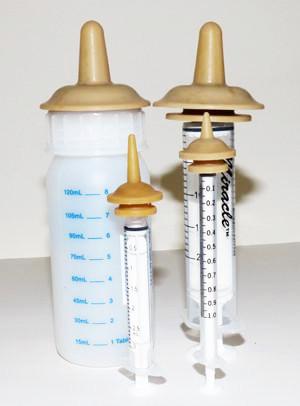
- Luer Lock Syringe 10 or 20 cc (will attach to feeding tube)
- 3cc - Premature kittens
- 3.5fr Feeding Tube - Premature kittens
- 5fr Feeding Tube – 1 to 6 Wks
- 8fr Feeding Tube – 6 to 10 Wks (Older kittens can chew tubes in half, use if emergency)
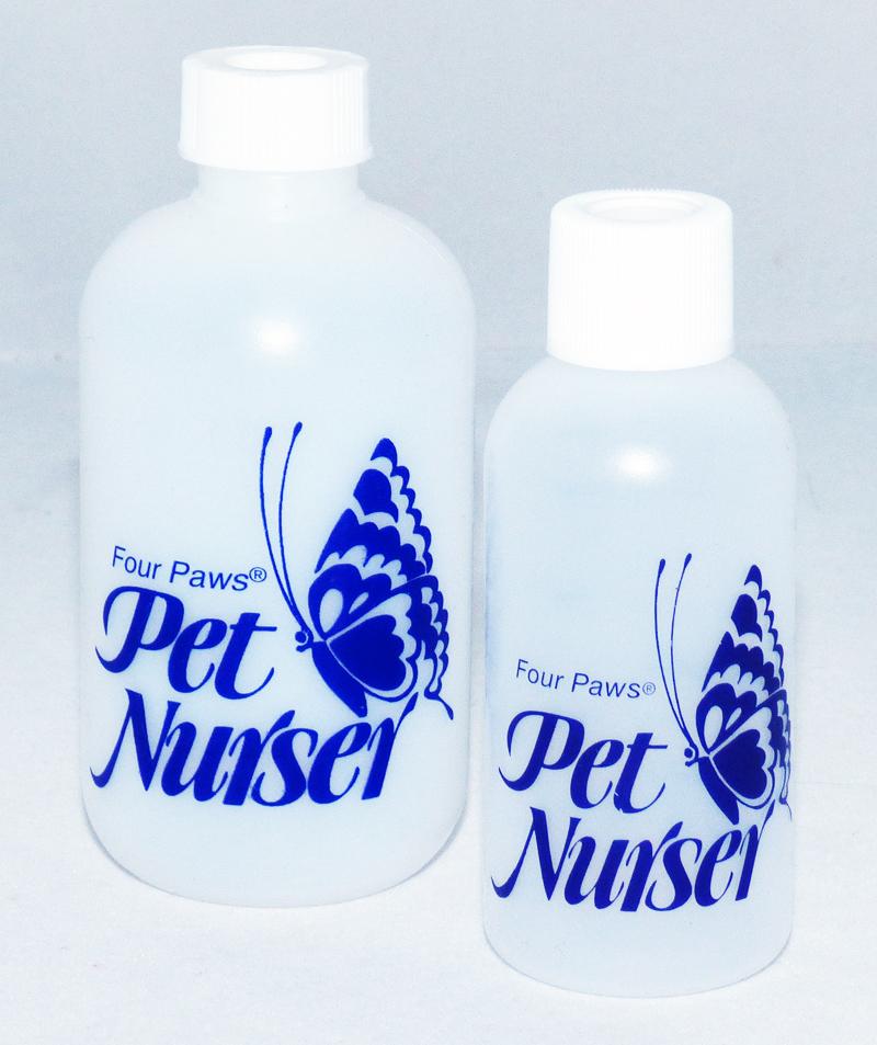
Bottle Feeding Supplies
- Bottles
- PetAg - 4 oz.
- Tighen tid to control milk flow.
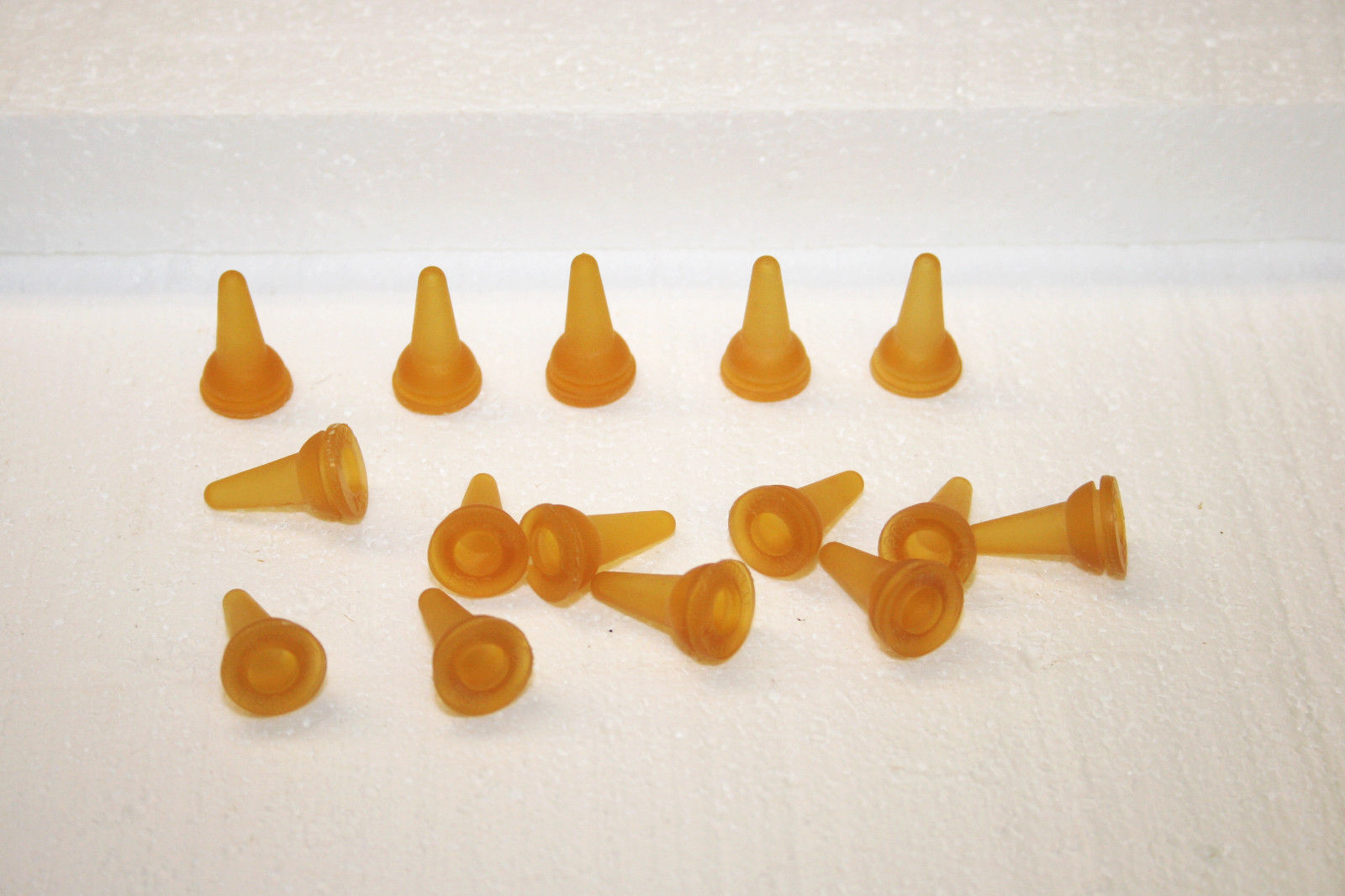
- Nipples
- PetAg Ready-to-Feed Nipple
- Use razor blade, shave small increments until desired hole size
- Milk
- Zoologic Milk Matrix 33/40
- Only use KMR in emergency, replace asap.
- Formula Mixer Bottle